Upper Thigh Muscles Ct Anatomy : Automated Assessment Of Regional Muscle Volume And Hypertrophy Using Mri Scientific Reports - Thin ct that runs outside of upper leg.. Each compartment is composed of muscles, neurovascular structures, and intermuscular fascia. Anatomical structures of the lower limb (hip, thigh, knee, leg, ankle and foot) and specific regions (compartment of the lower limb) are visible on dynamic labeled images. One further muscle of the anterior knee is the small articularis genus muscle, it is occasionally is blended with vastus intermedius. Lesser trochanter of the femur. Abdominal computed tomography (ct) is a type of medical imaging procedure used to diagnose and monitor internal stomach issues, like cancer, bowel obstruction, and abdominal pain.
Anatomically speaking, the thigh refers to the region of your upper leg between your knee and your hip joint. Diagnosis not applicable diagnosis not applicable. This mri brain cross sectional anatomy tool is absolutely free to use. The vastus lateralis is a muscle located on the lateral, or outside, part of your thigh. Upper thigh cross sectional anatomy :
Presentation1 Pptx Radiological Anatomy Of The Thigh And Leg from image.slidesharecdn.com You may also find transversus abdominis, iliopsoas, gluteus medius, pectineus, adductor longus. Also called the thigh bone, this is the longest bone in the body.it. Abdominal computed tomography (ct) is a type of medical imaging procedure used to diagnose and monitor internal stomach issues, like cancer, bowel obstruction, and abdominal pain. Muscles are named according to their shape, location, or a combination. Upper two thirds of the medial margin and proximal margin of the patella, medial condyle of the tibia, and investing deep fascia of the leg with the tendons of vastus intermedius, lateralis, and rectus, and through the patellar ligament onto the front of the tibial tuberosity. The iliacus muscle is part of a major trio of muscles in each hip joint also known as the iliopsoas —the iliacus muscle, the psoas major muscle, and the psoas minor muscle, that. There are five muscles in total, four of which form the powerful quadriceps muscle. There are five muscles in the anterior thigh compartment:
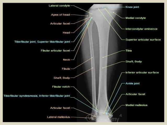
Anatomical structures of the lower limb (hip, thigh, knee, leg, ankle and foot) and specific regions (compartment of the lower limb) are visible on dynamic labeled images.
Learn vocabulary, terms, and more with flashcards, games, and other study tools. Muscle anatomy posterior 12 photos of the muscle anatomy posterior knee muscle anatomy posterior, muscle anatomy posterior view, posterior forearm muscle anatomy, posterior muscle anatomy chart, posterior thigh muscle anatomy ct, human muscles, knee muscle anatomy posterior, muscle anatomy posterior view, posterior. Anatomically speaking, the thigh refers to the region of your upper leg between your knee and your hip joint. Upper thigh cross sectional anatomy : Their origins and insertions are difficult to remember, and they are best considered as parts of general functional groups. Sartorius, and the four quadriceps muscles; In this image, you will find rectus abdominis, external oblique, inguinal ligament, tensor fascia lata, gracilis, sartorius, rectus femoris, the iliotibial band in it. Thin ct that runs outside of upper leg. Upper leg numbness, thigh weakness, thigh pain from overuse. Lesser trochanter of the femur. Muscles of upper back 12 photos of the muscles of upper back map of upper back muscles, muscles of the upper back and chest, origin and insertion of upper back muscles, superficial muscles of the upper back, tight muscles of the upper back and neck, human muscles, map of upper back muscles, muscles of the … Like the biceps brachii in the arm, the biceps femoris muscle has two heads. Each compartment is composed of muscles, neurovascular structures, and intermuscular fascia.
Iliac fossa for iliacus and lumbar spine for psoas muscles. When the leg muscles contract, it results in the pulling of muscle normally. Lesser trochanter of the femur. Anatomical structures of the lower limb (hip, thigh, knee, leg, ankle and foot) and specific regions (compartment of the lower limb) are visible on dynamic labeled images. Muscle anatomy posterior 12 photos of the muscle anatomy posterior knee muscle anatomy posterior, muscle anatomy posterior view, posterior forearm muscle anatomy, posterior muscle anatomy chart, posterior thigh muscle anatomy ct, human muscles, knee muscle anatomy posterior, muscle anatomy posterior view, posterior.
Lower Extremity Mri Anatomical Atlas from www.imaios.com Upper leg numbness, thigh weakness, thigh pain from overuse. Learn vocabulary, terms, and more with flashcards, games, and other study tools. Upper two thirds of the medial margin and proximal margin of the patella, medial condyle of the tibia, and investing deep fascia of the leg with the tendons of vastus intermedius, lateralis, and rectus, and through the patellar ligament onto the front of the tibial tuberosity. The thigh is the area between the hip and the knee joint. There are five muscles in the anterior thigh compartment: The muscles located within the posterior compartment of the thigh are the biceps femoris, semitendinosus and semimembranosus. Muscles are named according to their shape, location, or a combination. Groin muscles have adductors which are fan like structures present in the upper thigh region.
On the anterior side, the most prominent of the muscles are the sartorius muscle and the four muscles that make up quadriceps muscle group (the quads.)
The vastus laterails works with the other quad muscles to help extend your knee joint. Lesser trochanter of the femur. Muscles of upper back 12 photos of the muscles of upper back map of upper back muscles, muscles of the upper back and chest, origin and insertion of upper back muscles, superficial muscles of the upper back, tight muscles of the upper back and neck, human muscles, map of upper back muscles, muscles of the … Groin muscles have adductors which are fan like structures present in the upper thigh region. Muscle anatomy posterior 12 photos of the muscle anatomy posterior knee muscle anatomy posterior, muscle anatomy posterior view, posterior forearm muscle anatomy, posterior muscle anatomy chart, posterior thigh muscle anatomy ct, human muscles, knee muscle anatomy posterior, muscle anatomy posterior view, posterior. Start studying upper leg muscles. The thigh is the area between the hip and the knee joint. Online mri & ct sectional anatomy kenneth k. The muscles of the lower limb are numerous and complex. Also called the thigh bone, this is the longest bone in the body.it. Muscles of the lower limb; Case contributed by dr roberto schubert. You may also find transversus abdominis, iliopsoas, gluteus medius, pectineus, adductor longus.
Radiographers suggest an abdominal ct scan to look for the following: Anatomically speaking, the thigh refers to the region of your upper leg between your knee and your hip joint. Two (iliacus and psoas) muscles are deep in the abdominal cavity and superficial distal to the inguinal ligament and medial to the sartorius muscle. Related posts of muscle anatomy of the thigh muscles of upper back. 2, vastus medialis & intermedius muscles.
Fascial Compartments Of Thigh Wikipedia from upload.wikimedia.org The muscles of the anterior compartment of the thigh are located anterior to the femur. Start studying upper leg muscles. There are five muscles in the anterior thigh compartment: The majority of muscles in the leg are considered long muscles, in that they stretch great distances. Essentials of human anatomy and physiology 12th edition elaine n. The four muscles all extend the lower leg. These images are arranged in radiographic view. One further muscle of the anterior knee is the small articularis genus muscle, it is occasionally is blended with vastus intermedius.
These images are arranged in radiographic view.
On the anterior side, the most prominent of the muscles are the sartorius muscle and the four muscles that make up quadriceps muscle group (the quads.) Related posts of muscle anatomy of the thigh muscles of upper back. Lesser trochanter of the femur. The rectus femoris is located in the center of the thigh, while the vastus medialis is in the middle of the said body part. Related posts of upper anterior muscle anatomy muscle anatomy posterior. Cross sectional anatomy of the hip : Anatomical structures of the lower limb (hip, thigh, knee, leg, ankle and foot) and specific regions (compartment of the lower limb) are visible on dynamic labeled images. The majority of muscles in the leg are considered long muscles, in that they stretch great distances. Muscles are named according to their shape, location, or a combination. The muscles located within the posterior compartment of the thigh are the biceps femoris, semitendinosus and semimembranosus. There are five muscles in total, four of which form the powerful quadriceps muscle. Meanwhile, the vastus lateralis is on the side of the thigh, while the vastus intermedius is hidden below the rectus femoris(5). You may also find transversus abdominis, iliopsoas, gluteus medius, pectineus, adductor longus.
Welcome to online mri & ct sectional anatomy upper thigh anatomy. The thigh extends from the superior margin of the subtrochanteric region through the distal femoral metadiaphysis.


Posting Komentar
0 Komentar